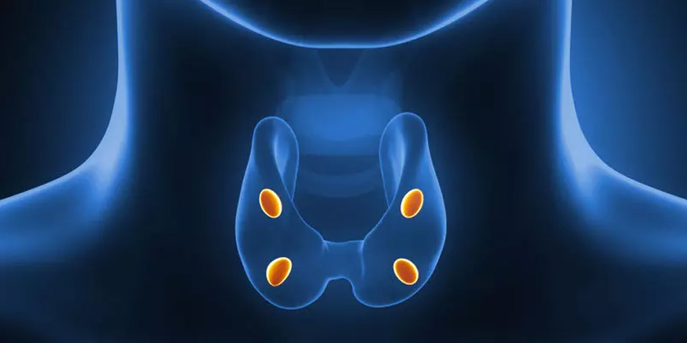
Parathyroid glands are glands located just behind the thyroid gland and secrete parathyroid hormone. There are 4 parathyroid glands, usually 4 mm in size. The parathyroid glands manage calcium metabolism in the body.
Hyperparathyroidism (High Parathormone)
Excessive secretion of parathyroid hormone for any reason is called hyperparathyroidism. There are three types of hyperparathyroidism: primary, secondary and tertiary. While surgical and medical treatments can be applied in secondary and tertiary hyperparathyroidism, the treatment of primary hyperparathyroidism is surgery. Patients who do not want to have surgery or who cannot have surgery due to the risks of surgery can be treated with ethanol ablation.
Primary Hyperparathyroidism (High Parathormone – Parathyroid Adenoma):
Overactivity of one or, rarely, more than one parathyroid glands is called primary hyperparathyroidism. The parathyroid gland grows and overworks, so parathyroid hormone (PTH-Parathormone) is secreted a lot. When PTH is secreted too much, calcium dissolution from the bone increases. If this process continues, there will be osteoporosis, bone pain, and blood-filled cavities called “brown tumors” appear in the bones and bone fractures occur.
Excess blood calcium causes kidney stones and kidney damage, ulcers and gastritis in the stomach and duodenum. In addition, constipation, nausea, muscle weakness, hypertension and psychiatric diseases (such as depression, mood disorders) occur in these patients.
Surgical treatment is parathyroidectomy, that is, removal of the parathyroid gland. Patients who do not want to have surgery or who cannot have surgery due to the risks of surgery can be treated with ethanol ablation.
Secondary hyperparathyroidism is the stimulation of the parathyroid glands due to external factors, increasing the production of PTH and undergoing changes. It is most commonly seen in patients with chronic renal failure. Diagnosis can be made with decreased ionized calcium, increased phosphorus and increased PTH levels in the blood. There are basically two treatment approaches in SHPT patients: medical (medicated) and surgical.
Tertiary hyperparathyroidism develops as a result of the parathyroid glands gaining autonomy after successful kidney transplantation in patients with long-term chronic renal failure of secondary hyperparathyroidism. The result is autonomously elevated PTH concentrations associated with elevated calcium levels.
Clinical Symptoms of Primary Hyperparathyroidism:
Bone pain, osteoporosis (bone loss) and bone fractures, kidney stones, nausea, ulcers, constipation, muscle weakness, early fatigue, weakness, difficulty concentrating, memory problems are seen. Cardiac findings include elevated blood pressure and bradycardia (reduction in pulse rate), and left ventricular hypertrophy.
Laboratory Tests Required for Diagnosis:
Blood calcium level and albumin level are measured together or ionized calcium level is measured. Normal blood calcium is usually between 8.5-10.5 mg/dl and ionized calcium is between 1.13-1.32 mmol/L. The blood calcium (Ca) level is related to the albumin level. If the albumin level is low, the calcium level is measured low at its true value. Therefore, the corrected calcium value is calculated.
Corrected Calcium = Measured total Ca + [0.8 x (4.0 – albumin level)]
The normal value of albumin is between 3.5-5.5 mg/dl. If the albumin value is lower than normal, the corrected calcium value is actually higher. It is important to calculate the corrected calcium value in the elderly and those with additional diseases.
When the calcium level is found to be at least twice as high, the parathyroid hormone level is checked. If the serum calcium or ionized calcium and parathormone levels are high, the diagnosis of primary hyperparathyroidism is made.
In order to exclude rare diseases such as familial benign hypercalcemia disease, 24-hour urinary calcium excretion is evaluated. In these patients, 24-hour urinary calcium excretion is lower than normal.
Hypercalcemia due to lithium (a drug given to manic-depressive patients) should also be considered in the differential diagnosis.
Finally, since vitamin D is low, parathormone may be secreted too much to increase it. Vitamin D value in the blood should be checked, and parathormone and calcium should be checked again after vitamin D support therapy is given in patients with low blood levels.
With excessive secretion of parathormone, calcium levels increase, phosphate levels decrease, serum chlorine values may be normal or high.
Imaging Diagnostic Methods:
Neck Ultrasonography: Generally, parathyroid gland adenomas are diagnosed by ultrasonography. An adenoma behind the thyroid gland is seen on ultrasound in 75-80% of patients.
Parathyroid Scintigraphy: Scintigraphy is not necessary in every case. The sensitivity of parathyroid scintigraphy in recognizing parathyroid adenomas is between 60-90%.
Computed tomography (CT) or neck magnetic resonance imaging (MRI) can be performed in cases that do not display adenoma together with ultrasonography and scintigraphy .
Non-surgical treatment of parathyroid adenoma (Ethanol Ablation )
The method we call ethanol ablation can be applied in patients with primary hyperparathyroidism in which the parathyroid gland is enlarged, as well as in other hyperparathyroidisms with high calcium values.
Although it is a procedure that requires experience since the parathyroid gland is deep in the neck, ethanol is injected into the parathyroid gland only after a needle reaches the parathyroid gland correctly. Ethanol causes tissue damage, reducing the gland’s hormone secretion. A single injection is usually sufficient for this procedure.
An experienced interventional radiologist can easily perform the treatment by injecting the appropriate dose. Usually, the calcium levels of the patients who are treated after a single injection return to normal on the same day. In our clinic, we can follow the blood calcium values every 3 months after the treatment and plan additional procedures if necessary. Parathyroid adenoma treatment with ethanol ablation without surgery is a low-risk and easy treatment, so it can be easily reapplied if there is not enough calcium reduction. If you want to review the results of ethanol ablation, you can read the following articles(1-5).
Scientific References
- Cappelli C et al. Modified percutaneous ethanol injection of parathyroid adenoma in primary hyperparathyroidism. QJM. 2008 Aug;101(8):657-62. doi: 10.1093/qjmed/hcn062. Epub 2008 May 22.
- Vergès BL et al. Results of ultrasonically guided percutaneous ethanol injection into parathyroid adenomas in primary hyperparathyroidism. Acta Endocrinol (Copenh). 1993 Nov;129(5):381-7.
- Alherabi AZ et al. Percutaneous ultrasound-uided alcohol ablation of solitary parathyroid adenoma in a patientwith primary hyperparathyroidism. Am J Otolaryngol. 2015 Sep-Oct;36(5):701-3. doi: 10.1016/j.amjoto.2015.04.006. Epub 2015 Apr 15.
- Chen HH et al. Effects of percutaneous ethanol injection therapy on subsequent parathyroidectomy . 2008 Aug;196(2):155-9. doi: 10.1016/j.amjsurg.2007.06.037. Epub 2008 May 29.
- Nakamura M et al. Effects of percutaneous ethanol injection therapy on subsequentsurgical parathyroidectomy
Prof. Dr. Mehmet Mahir Atasoy
Interventional Radiology


