
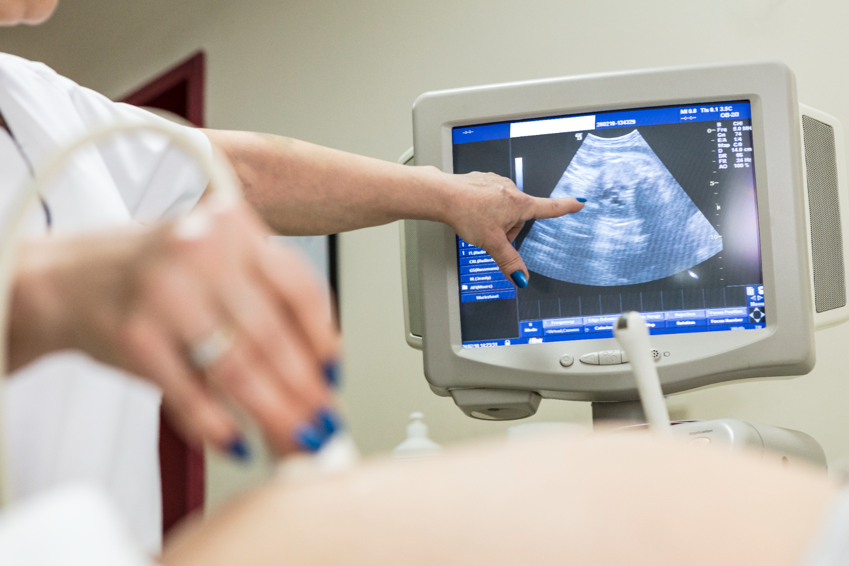

Breast Ultrasonography
Breast ultrasonography is a safe and non-radiating imaging method that evaluates the internal structure of breast tissue using high-frequency sound waves. It provides great advantages in early diagnosis, particularly in young women, those with dense breast tissue, and when used together with mammography.
Dr. Atasoy Clinic provides breast ultrasonography service with advanced-technology ultrasound equipment in the field of breast health, ensuring high diagnostic accuracy and patient comfort.
What is Breast Ultrasonography and What is Its Purpose?
Breast ultrasound allows sound waves to be transmitted to tissue through a special gel applied to the skin. After these waves strike the tissues, they are reflected back and converted into images by the computer.
This way, abnormalities such as masses, cysts, fibrocystic changes, inflammation, or tumors in the breast are easily detected.
Because it does not contain radiation, it can be safely used in all age groups, pregnant women, and nursing mothers.
Additionally, breast ultrasound is used as a guide during breast biopsy, drainage, or ablation procedures.
When Should It Be Performed?
Breast ultrasonography is one of the screening methods every woman should undergo regularly.
However, in certain situations, more frequent performance for diagnostic purposes is recommended.
The main situations where breast ultrasound is recommended are:
-
Family history of breast cancer
-
Palpable swelling, mass, or pain
-
Nipple discharge
-
Skin dimpling, color changes, or asymmetry
-
Suspicious findings on mammography
-
Control purposes in patients with silicone implants
-
Annual screening for women under 40
At Dr. Atasoy Clinic, ultrasound screening frequency is determined according to individual risk factors.
How Is the Procedure Performed?
Breast ultrasound is completely painless, comfortable, and quick.
No preparation is required; prior to the procedure, no special diet or medication discontinuation is necessary.
Procedure steps are as follows:
-
The patient lies on her back with arms behind her head.
-
Special ultrasound gel is applied to the area to be examined.
-
The breast tissue is scanned with a high-resolution probe.
-
If necessary, lymph node regions of both breasts are also evaluated.
-
Images are recorded and findings are interpreted immediately by the specialist doctor.
The entire procedure takes approximately 10-15 minutes and the patient can return to normal daily activities immediately after the exam.
Advantages of Breast Ultrasound
-
Contains no radiation; completely safe.
-
Painless and non-invasive.
-
High success in early diagnosis.
-
Cystic and solid lesions can be differentiated.
-
Ideal for pregnant women and young women.
-
Can be used as a guiding tool during biopsy or ablation.
In individuals with dense breast tissue, diagnostic accuracy significantly increases when used together with mammography.
What is the Difference from Mammography?
| Feature | Breast Ultrasound | Mammography |
| Method | Sound waves | X-rays |
| Radiation | None | Present |
| Mass Detection | Can differentiate cystic and solid lesions | Shows microcalcifications |
| Age Group | Ideal for under 40 | Recommended for over 40 |
| Use in Pregnancy | Safe | Not recommended |
| Pain / Compression | None | May be mild |
These two methods complement each other. Mammography is used for general screening while ultrasound is used for detailed examination and mass characterization.
What Lesions Can Be Detected?
Breast ultrasound can visualize many different pathologies:
-
Simple or complicated breast cysts
-
Fibroadenomas (benign tumors)
-
Breast abscess or inflammatory processes
-
Fat necrosis and post-trauma changes
-
Small masses without microcalcifications
-
Irregular lesions suspicious for cancer
If these findings are detected early, the treatment process becomes much easier and more effective.
Interpretation of Results
At Dr. Atasoy Clinic, all breast ultrasound results are reported according to the Breast Imaging Reporting and Data System (BI-RADS) classification.
BI-RADS is an international system that determines the risk level of lesions:
| BI-RADS Category | Description |
| 1 | Normal, no pathology |
| 2 | Benign findings |
| 3 | Probably benign, follow-up in 6 months |
| 4 | Suspicious finding, biopsy recommended |
| 5 | High likelihood of malignancy, further evaluation |
| 6 | Malignancy confirmed by biopsy |
This system clearly establishes follow-up and treatment planning.
Breast Ultrasound Experience at Dr. Atasoy Clinic
Dr. Atasoy Clinic places great importance on early diagnosis in breast health.
Thanks to the high-resolution ultrasound systems used, even lesions of millimeter scale can be clearly visualized.
All examinations are performed by experienced radiology specialists and the patient is informed immediately.
If needed, a biopsy plan or interventional procedure is developed in the same session.
The clinic's approach is based on the principle of "detect early, treat early."
The goal is for every woman to understand her own breast tissue, develop regular check-up habits, and be monitored according to her individual risk.
