
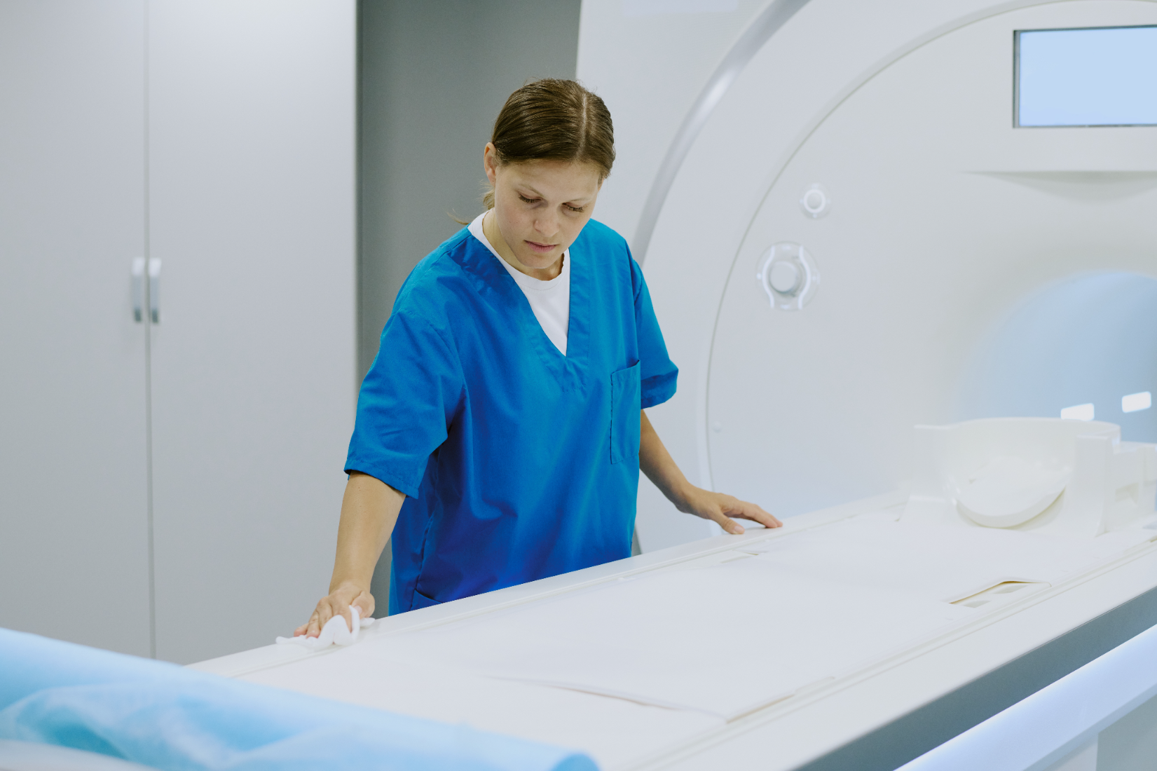

Breast Magnetic Resonance Imaging (MRI)
Breast magnetic resonance imaging (MRI) is one of the most advanced imaging methods for comprehensive evaluation of breast tissue. It plays a very important role in diagnosis and follow-up, particularly in high-risk patients, due to its lack of radiation, high-resolution detail, and ability to detect even early-stage breast cancers.
Dr. Atasoy Clinic performs breast MRI examinations with high accuracy using modern MRI technology and special breast coils, providing patients with significant diagnostic advantages for early detection.
What is Breast MRI and How Does It Work?
Breast MRI is a method that creates three-dimensional and detailed images of breast tissue using powerful magnetic fields and radio waves.
During imaging, contrast agent (gadolinium) is typically used. This substance is administered intravenously and enhances the blood supply characteristics of tumor tissue. This way, the differences between benign and malignant lesions become clearly visible.
Breast MRI is not merely a screening method; it is also used in treatment planning, biopsy guidance, and post-operative follow-up procedures.
When Is Breast MRI Performed?
Breast MRI is generally used when mammography or ultrasound results are insufficient or unclear.
However, in certain special situations, it is recommended as the first choice.
The main situations where breast MRI is recommended are:
-
Individuals with family history of breast cancer and high genetic risk (BRCA1, BRCA2 positive)
-
Patients with dense breast tissue
-
Differentiation of suspicious lesions detected by mammography or ultrasound
-
Post-operative surveillance or suspicion of recurrence
-
Evaluation of silicone integrity in patients with implants
-
Monitoring treatment response after chemotherapy or radiotherapy
-
Metastatic workup or suspicion of multiple lesions
At Dr. Atasoy Clinic, the decision for MRI is individually evaluated in light of the patient's clinical history and other imaging results.
How Is the Procedure Performed?
Breast MRI is completely painless and safe.
No special preparation is needed before the procedure; however, metal jewelry and accessories should be removed.
For patients with contrast allergy, testing is performed in advance.
Procedure steps are as follows:
-
The patient is placed prone on a special breast MRI table.
-
Both breasts are positioned within specially designed MRI coils.
-
If needed, contrast agent is administered intravenously.
-
During imaging lasting 15-25 minutes on average, the patient must remain still.
-
Images are obtained and evaluated in detail by the radiologist.
The patient experiences no pain during the procedure, only hears the magnetic sounds of the machine.
For patients with claustrophobia, mild sedation can be provided.
Advantages of Breast MRI
-
Has the highest tissue resolution of any breast imaging method.
-
Contains no radiation; completely safe.
-
Most sensitive method for differentiating benign and malignant lesions.
-
Ideal for evaluating silicone integrity in augmented breasts.
-
Detects multifocal tumors and occult lesions.
-
Can detect recurrence early in the post-treatment period.
-
Guides combined treatment planning (surgery + radiotherapy).
These advantages have made breast MRI indispensable, particularly for high-risk women.
Comparison with Other Imaging Methods
| Feature | Breast MRI | Mammography | Ultrasound |
| Radiation | None | Present | None |
| Image Detail | Very high | Moderate | Moderate |
| Tissue Density Effect | Unaffected | Reduces visibility | Affected |
| Implant Evaluation | Excellent | Limited | Limited |
| Multiple Lesion Detection | High success | Moderate | Moderate |
| Application Area | Diagnosis, screening, treatment follow-up | Screening | Initial evaluation |
This table clearly shows that breast MRI used together with other methods increases diagnostic accuracy.
Interpretation of Results
At Dr. Atasoy Clinic, breast MRI results are reported according to BI-RADS MRI classification.
Each finding is evaluated based on its contrast uptake characteristics and morphological appearance.
This classification provides diagnostic certainty and guides further diagnostic planning (biopsy, ablation, etc.) if necessary.
Additionally, during the reporting process, previous ultrasound or mammography findings are examined comparatively.
This comprehensive approach minimizes false positive results.
Breast MRI Experience at Dr. Atasoy Clinic
Dr. Atasoy Clinic uses high-resolution 1.5T and 3T devices, special breast coils, and advanced imaging software for breast MRI imaging.
For each patient, the imaging protocol is planned according to personal risk status and complaint.
The reporting process is performed by experienced radiologists according to international criteria.
If needed, biopsy planning or treatment guidance is also provided in the same session.
The clinic's approach is not only to detect disease; it is designed to identify risks in early stages, increase treatment success, and provide patients with a safe follow-up process.
